

Cervical cancer is the fourth most common malignant tumor among women worldwide, causing over 300000 deaths annually. However, the identification of cervical cancer stem cells is still unclear. A small group of cells with unlimited proliferation potential in tumors that can reconstruct tumor development are called cancer stem cells, also known as tumor initiating cells. It is a small specialized subpopulation of malignant cells that maintains a relatively stable proportion in tumors through self-renewal and differentiation, and is tolerant to therapies such as radiotherapy and chemotherapy. The origin of tumor stem cells is still unclear.
In order to explore the origin of tumor stem cells, some studies suggest that they originate from differentiated non stem cells, which acquire stem cell like features after malignant transformation. In order to explore the transformation process of malignant tumor stem cells in cervical cancer patients and the cellular composition and interactions of the cervical cancer microenvironment, the author of this article combined multiple omics techniques to provide a detailed description of cellular characteristics.

On May 22, 2023, the Cancer Biology Research Center of Tongji Hospital affiliated with Tongji Medical College of Huazhong University of Science and Technology published an article titled "Identification of cervical cancer stem cells using single cell transcriptomes of normal Cervix" in the eBioMedicine journal, Research paper on cervical premalignant lesions and cervical cancer. This provides more research content for the microenvironment of cervical precancerous and malignant lesions.
This article performed mRNA sequencing on approximately 122400 single cells from 20 cervical biopsy specimens (including 5 healthy controls, 4 high-grade cervical intraepithelial lesions, 5 cervical microinvasive carcinoma, and 6 cervical squamous cell carcinoma), and validated the bioinformatics results on cervical cancer tissue microarray (TMA) using multiple immunohistochemistry (mIHC).
The experiment identified cervical cancer stem cells and revealed the functional changes of cervical stem cells during malignant transformation. The primitive non malignant stem cell characteristics (characterized by high proliferation) gradually weaken, while the tumor stem cell characteristics (characterized by epithelial mesenchymal transition and invasion) are enhanced. Our TMA chip's mIHC results confirm the presence of cancer stem cell like cells and indicate that this cell subset is associated with tumor recurrence. In addition, we also investigated the malignant and immune cell heterogeneity in the cervical multicellular microenvironment structure at different stages of lesions. Moreover, an overall upregulation of interferon response in the cervical microenvironment was observed during the progression of lesions.
Experimental part
To verify the presence of cell populations with specific transcriptional features in cervical cancer tissues, The author used Tissue Cytometry technology to study the TMA chip of cervical cancer tissue. Under multiple staining labels, the tumor tissue was automatically identified and a microenvironment spatial analysis system was established. Not only did the presence of C2 subgroups in the tumor tissue be determined through precise single-cell recognition algorithms, but also the distribution of different cell populations in the tumor microenvironment was visualized through clustering analysis and annotation of data using transcriptome technology, in order to infer the interaction relationship between target cell populations.
The author of the article stated that the value of this publication lies in providing theoretical significance for understanding and treating tumor behaviors such as drug resistance and metastasis related to cervical cancer stem cells. In further experimental exploration in the future, Tissue Cytometry technology, as a precise quantitative analysis tool for tissue in situ, can help more researchers to more quickly and accurately verify data such as protein expression, cell distribution, and tissue-specific changes, laying a more solid foundation for the reliability of multi omics research techniques.
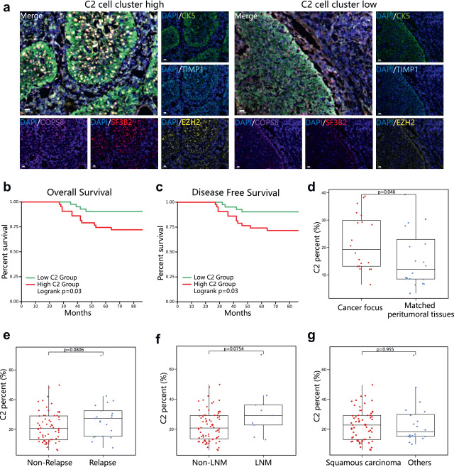
Figure 1 Verify the correlation between the percentage of high C2 cluster cells and poor prognosis in patients.
(a)Display immunofluorescence images of C2 cluster cells in cervical cancer tissue using multiple fluorescence immunohistochemistry staining (CK5, green; TIMP1, Blue color; EZH2, Yellow; SF3B2, red COPS8, Purple; PanCK CK5 is used to label cancer cells.
