MULTISPECTRAL CYTOMETERS

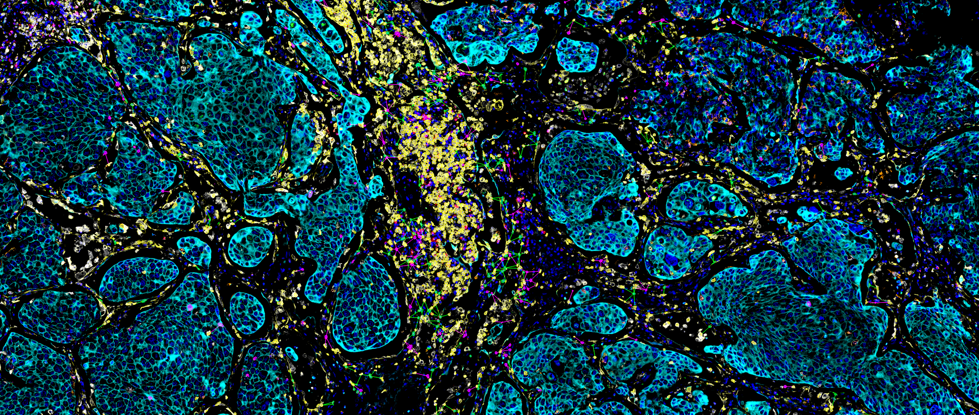
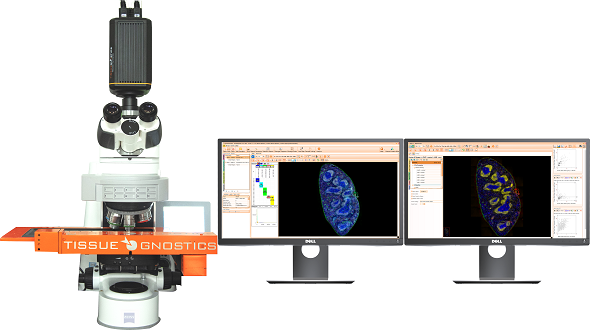
TissueFAXS Spectra enables multi-color panoramic continuous spectral imaging, multiplex fluorescence staining spectral unmixing, and the removal of autofluorescence from blood cells or samples. It overcomes the limitations of traditional multi-channel fluorescence imaging in terms of crosstalk and unmixing, which cannot accurately image and quantify multi-color samples. This allows for precise quantitative analysis of single cells in the tissue microenvironment in situ with multiple targets. Additionally, it features multi-level tissue image recognition and tissue-like flow analysis capabilities, accurately identifying single cells and specific structural regions (such as glands, tumor areas, blood vessels, bronchi, etc.) in complex tissues. It performs localization, qualitative, and quantitative analysis at multiple levels, including single cells, tissue structures, and cellular spatial information. This includes quantitative analysis of staining intensity and morphology of fluorescently labeled proteins, nucleic acids, and other components; sample cell subtype analysis and screening based on fluorescence labeling and morphological parameters; and quantification of cell-to-cell positional relationships and spatial distribution.

KEY FEATURES

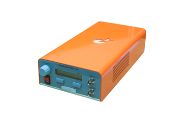
Independent 8-channel LED and laser hybrid light source

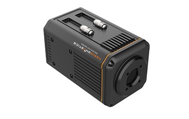
Fast spectral, full-spectrum continuous scanning and spectral splitting

Independent brightfield and spectral dual imaging module

Quickly view single-channel raw spectral calibration and splitting results

Customised independent spectral background deduction

Freely manage and call multiple spectral libraries

Multi-Spectral Technology
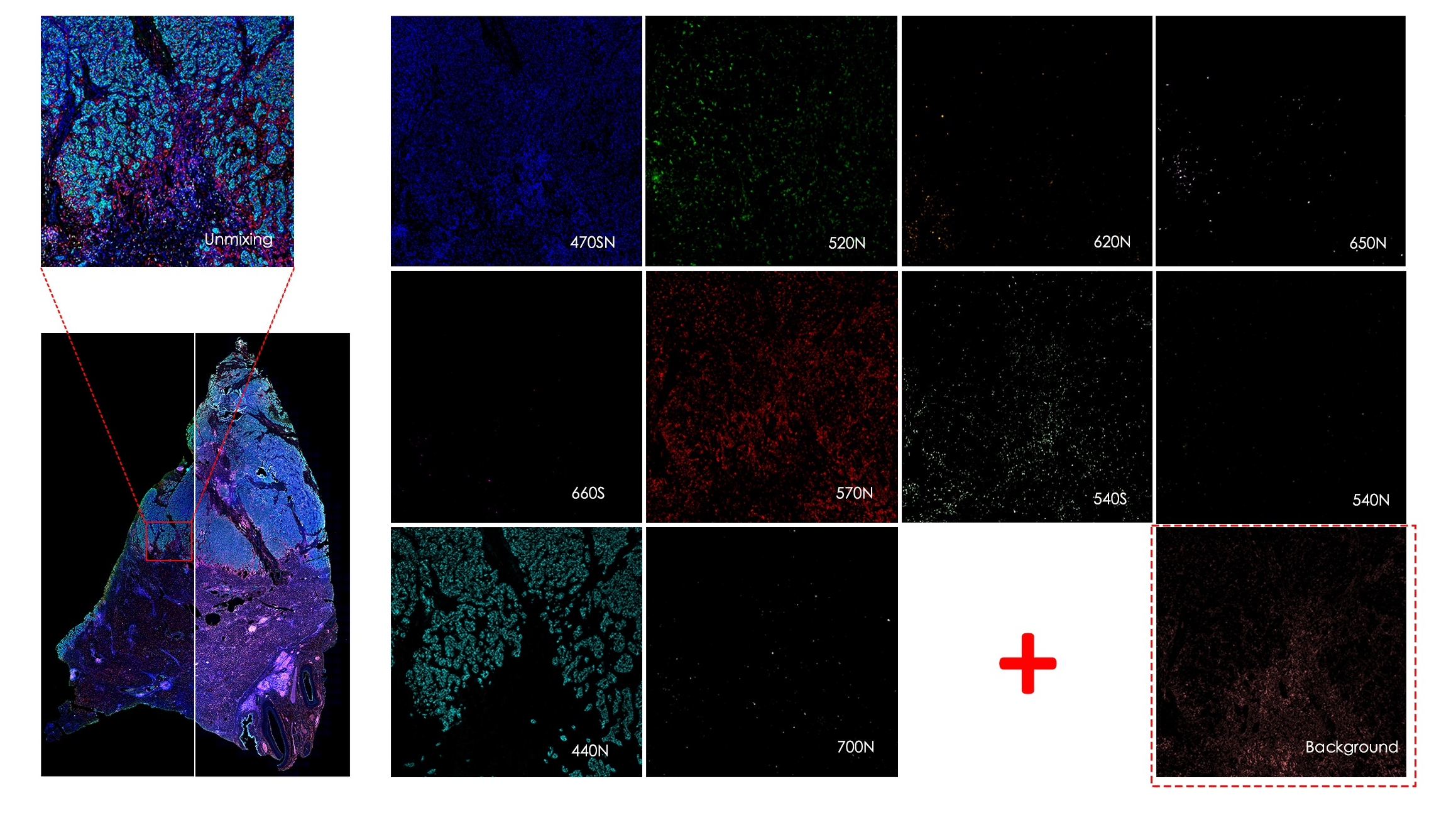
The TissueFAXS Spectra panoramic tissue multispectral imaging analysis system, through λ-stack multispectral imaging combined with spectral unmixing algorithms, clarifies which dyes and their proportions contribute to each pixel’s signal in the image. This technology resolves the issue of crosstalk between channels in multi-color labeling, providing more accurate signal intensity and morphological information for subsequent image quantification. TissueFAXS Spectra supports upgrades on other TissueFAXS series products (both upright and inverted systems).

Multispectral microscopy imaging is mainly applied in:
·Tumor microenvironment research
·Molecular marker research
·Spatial biology research
·Immunophenotyping analysis
·Immune infiltration analysis
High-throughput Cyc-IF staining and quantitative analysis techniques

High-throughput cyclic immunofluorescence(Cyc-IF) quantitative analysis technology constructs high-dimensional information images by performing multiple rounds of IF panoramic scanning on conventional sections, capable of detecting more than 60 antigen information. By using traditional IF and the process of fluorescein inactivation based on light and acid/alkali catalyzed oxidation, autofluorescence can be greatly reduced, and the signal-to-noise ratio is improved with the increase in staining rounds. Compared to the TSA staining method, where the non-linear coupling relationship between fluorescence intensity and protein expression prevents quantification of protein content through fluorescence signal intensity, the high-throughput Cyc-IF quantitative analysis method is simple in principle, fast in imaging speed, and has a large number of staining channels. It can also use TissueFAXS Cytometry quantitative analysis technology to determine single-cell protein expression levels based on staining fluorescence intensity and perform deep big data analysis on the quantity, morphology, location, and structure of any channel and any signal (cells, fluorescent probes, tumor tissues, nerve fibers, etc.).

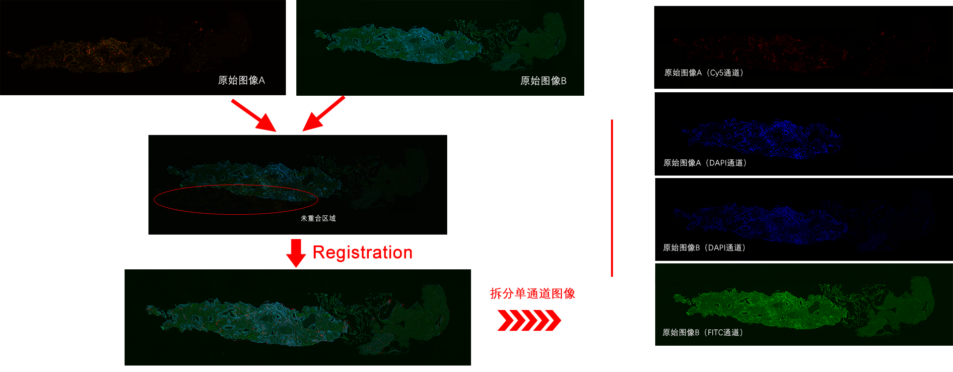
Using the Sample Registration function of StrataQuest software, target samples can be selected for overlay processing to obtain multi-round staining multi-channel overlay images centered on DAPI. By calculating the situation of DAPI nuclei, accurate quantitative analysis of nuclear, cytoplasmic, and membrane signals in each channel can be performed.


λ-stack imaging and spectral splitting of fluorescent samples

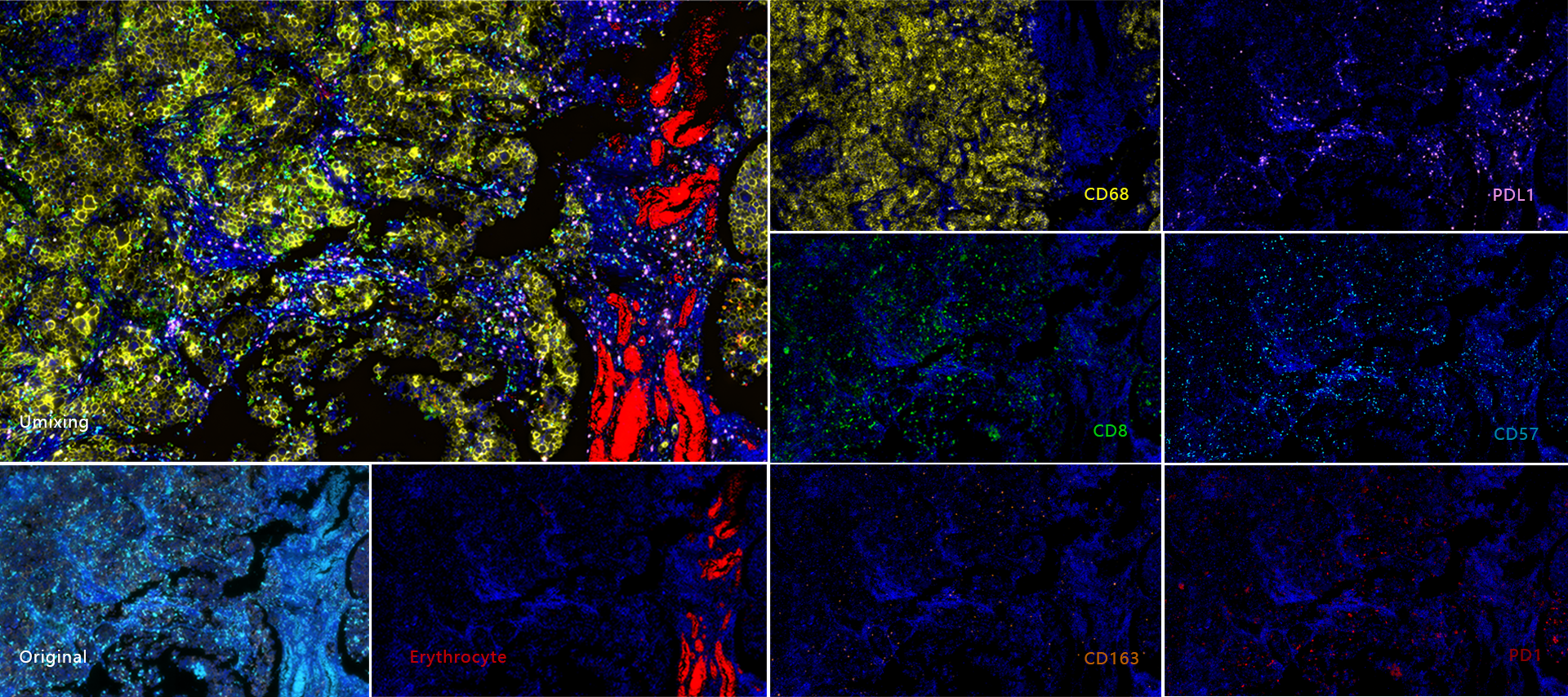
In the field of microscopic imaging, especially in the imaging of immunofluorescence samples, when there are too many labelled fluoresceins, the spectra of each fluorescein overlap. With traditional filter imaging, multiple fluorescent signals will be captured in a single channel, resulting in channel crosstalk and the inability to obtain a pure single-channel image.TissueFAXS Cytometry technology uses λ-stack multispectral imaging combined with spectral splitting algorithms to clearly identify which dyes and the proportion of dyes in each pixel in the image, thus solving the problem of crosstalk between channels when multi-colour labelling is performed. It solves the problem of colour crosstalk between channels during multi-colour labelling, thus providing more accurate signal intensity and morphological information for image quantification at a later stage.

Custom Spectrum Building and Dynamic Splitting

TissueFAXS Spectra library building and splitting mode adopts a flexible free combination strategy. In addition to splitting multiple staining signals based on relevant dyes and background spectra already in the spectral library, it can also acquire spectral data in real time for different autofluorescent backgrounds and add them to the splitting criteria automatically. For example, in samples with high autofluorescence background signals, blood cell background, collagen background, and multiple tissue background are deducted respectively. The number of target fluorescence spectra and the number of tissue background spectra in the spectral library can be added without upper limit, and multiple staining signals can be split within the spectral scanning range.

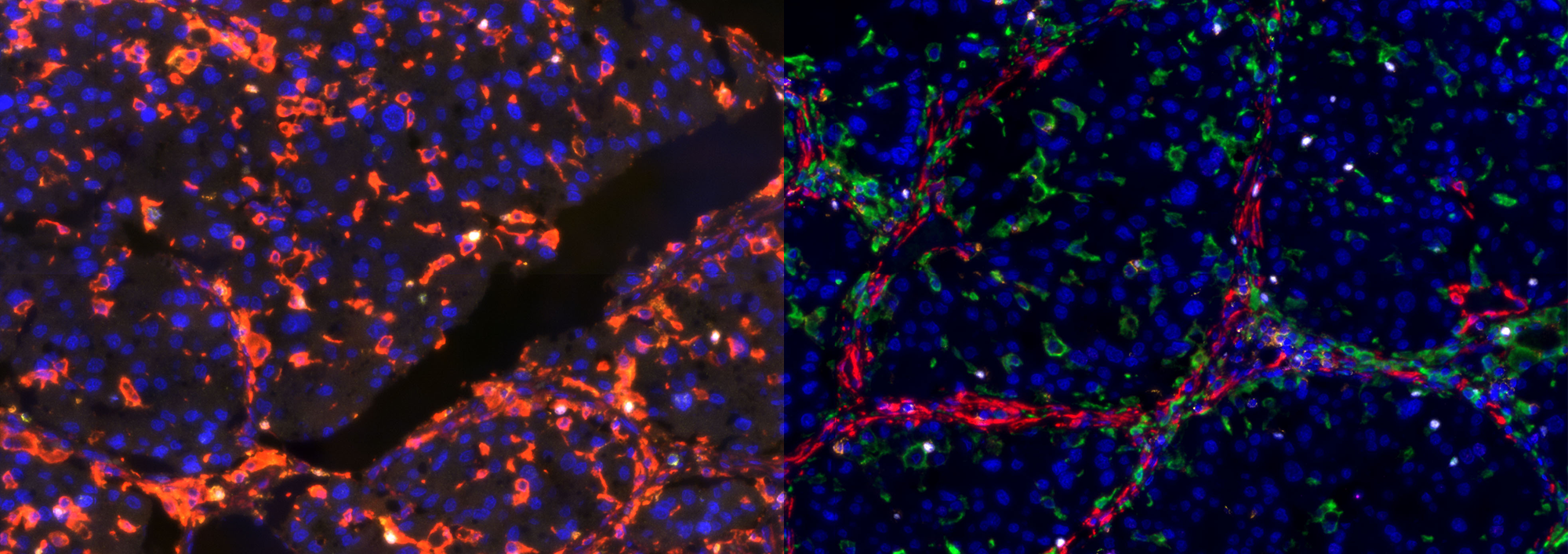
10+1-colour of liver tumour

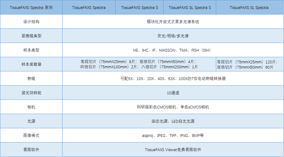
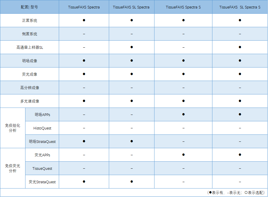
技术参数及配置