CONTEXTUAL TISSUE CYTOMETRY

The StrataQuest Tissue Flow Quantification Platform, integrated with HistoQuest/TissueQuest software, provides a solution for accurate quantitative analysis of tissues at the single-cell level in situ. The software is compatible with a wide range of tissue and cell samples, enabling a full range of in-situ quantitative analyses in different biological research fields, and exploring future needs for deeper analyses.

StrataQuest software can accurately identify complex tissues:

Molecular probe markers

Cellular sub-level structural markers 

Single-cell structural markers 

Specific tissue types 


KEY FEATURES

Automatic Recognition of Tissue Structures

Single cell recognition algorithm based on neural network and deep learning

AI Artificial Intelligence Tissue Traitised Data Mining

Spatial location distribution of cell subtypes

Multi-target tissue microenvironment protein-molecule interactions

Visualising multi-dimensional analysis of big data

Single cell recognition algorithm based on Deep Neural Network (DNN) and deep learning

In 2020, TissueGnostics developed a new generation of AI-based single cell nuclei recognition method based on the single cell nucleus recognition algorithm and combined with the deep neural network algorithm (DNN). Boundary identification eliminates the uncertainty caused by fuzzy boundaries on non-focal planes and improves the accuracy of nuclei/plasm/membrane morphological identification. (The left picture shows the conventional cell nucleus identification algorithm, and the right picture shows the single cell identification algorithm based on neural network (DNN) and deep learning)


Morphological recognition of specific tissue structures

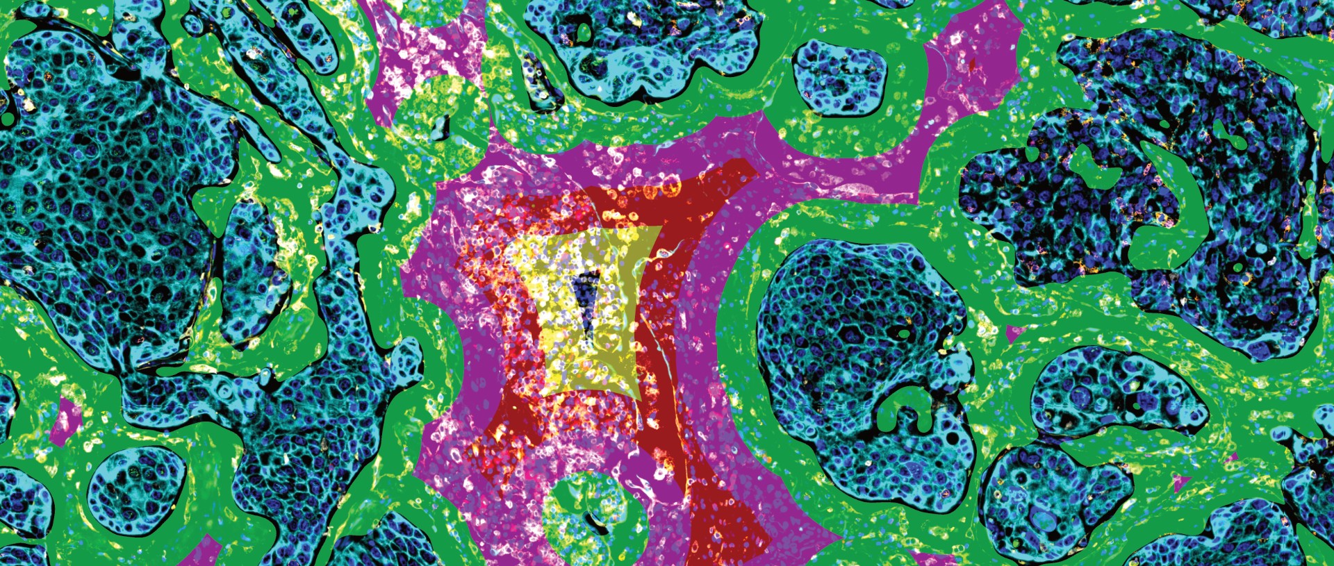
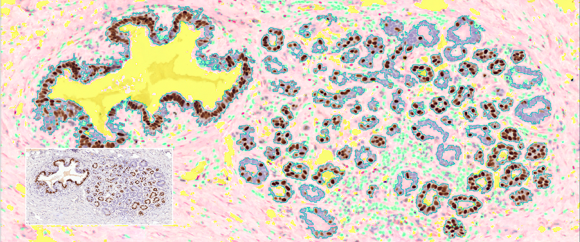
Specific tissue region identification solution for brightfield/fluorescent samples. For colour brightfield samples, automatic and independent nucleus/plasmid/membrane staining channels: 1. Using hematoxylin/DAPI staining channels, accurate identification of cell nuclei, followed by quantification of the distribution of nucleus/plasmid/membrane signal intensity and area morphology; 2. Using specific staining and tissue characteristics, identification of epithelial cells/mesenchymal regions/cavities, etc., and identification of tumours/glands/collagenous proliferation, etc., and quantitative analysis of the tissue type. and quantitative analysis. The solution can also be used for cell-tissue structure identification of HE-stained samples, making it possible to perform rapid staining + in-depth analysis of a large number of samples.