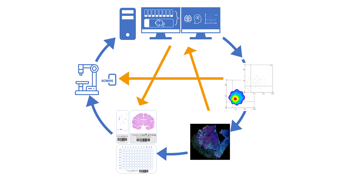
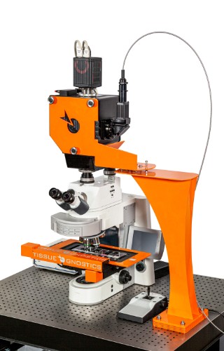
The TissueFAXS Q+ 2D/3D multidimensional panoramic tissue imaging quantitative analysis system acquires a large number of high-definition image samples at the histological level on a confocal plane. Starting from a panoramic imaging perspective, the system can obtain comprehensive data analysis results for the entire sample, including individual cells, specific structures, and spatial relationships. It supports panoramic scanning with objectives ranging from 2.5X to 100X oil immersion.
Traditional confocal devices are typically used for imaging specific cell structures or protein molecular markers stained within a single field of view but are unable to provide spatial location information of cells or specific structures across an entire slide. The TissueFAXS Q+ 2D/3D multidimensional panoramic tissue imaging quantitative analysis system, based on immunohistochemical staining techniques and panoramic stitching scanning imaging technology, effectively resolves the aforementioned issues, including the effect of weak photobleaching.


