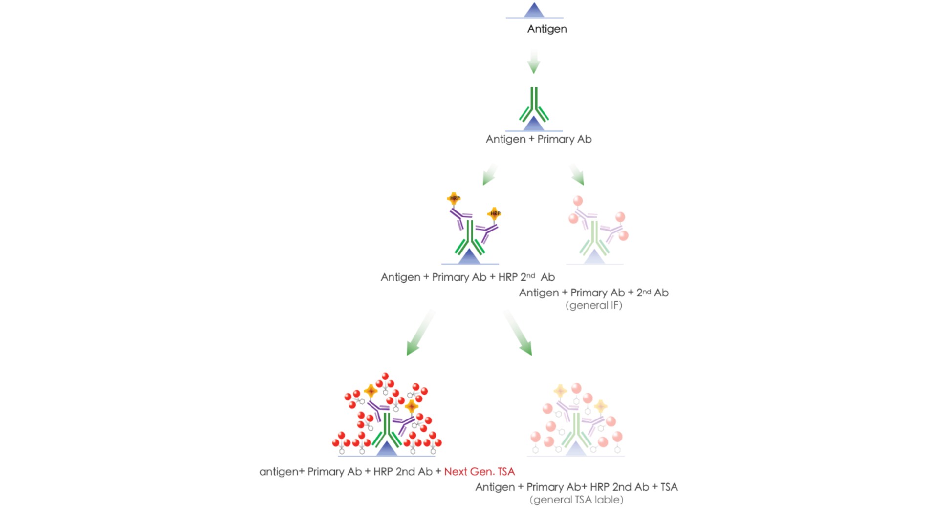
TSA Multiplex IHC Assay Kits

TissueGnostics provides cutting-edge technology—tissue cytometry and has become a leading brand in computational pathology. To lower the entry barriers of multiplex immunofluorescent staining (mIHC), TissueGnostics launched Tyramide Signal Amplification (TSA) mIHC Kits. TSA mIHC kits can amplify signals up to 100 folds and be adopted with TissueFAXS product lines (TissueFAXS Plus, Spectra, Q+), then be quantified by tissue cytometry. Certified protocols ensure your performance in multiplexed experiments.


Product features

Detection of low-abundance targets

Compatible with ICC,IHC,and FISH

Sensitivity up to 100-folds of conventional methods

Similar workflow to conventional ICC,IHC,and FISH

Use of less primary antibody

Simplified primary antibody panel design for multiplex IHC

Next Generation TSA Technology
Tyramide Signal Amplification is a highly sensitive method enabling the detection of low-abundance targets in fluorescent immunocytochemistry (ICC), immunohistochemistry (IHC), and in situ hybridization (FISH) applications.
 
TSA involves horseradish peroxidase (HRP) catalyzed tyramide deposition on and near a target protein or nucleic acid sequence in situ. In the presence of low concentrations of H2O2, HRP can convert a labeled tyramide substrate into a highly reactive form that can covalently bind to tyrosine residues on proteins at or near the HRP. This generates high-density tyramide labeling and is the reason for the exceptional sensitivity of this system. Tyramide can be labeled with fluorophores or hapten (such as biotin or DNP).
Advantages of Tyramide Signal Amplification
· Detection of low-abundance targets
· Compatible with ICC, IHC, and FISH
· Sensitivity up to 100-folds of conventional methods
· Similar workflow to conventional ICC, IHC, and FISH
· Use of less primary antibody
· Simplified primary antibody panel design for multiplex IHC
Multicolor labeling using Tyramide Amplification Kits
· No limitation on the choice of the primary antibodies
· Rapid FFPE antigen retrieval, only 5 mins in the microwave
· Next Generation TSA Technology:Amplify signals up to 100 folds
· Compatible with a conventional fluorescent microscope and TissueFAXS Spectra system
Multicolor imaging system: TissueFAXS Spectra
· Spectral unmixing overcomes the cross-talk between neighboring emission wavelengths.
· A wavelength resolution of 10 nm unmixes dyes with high spectral overlap
· Unmix up to 11 colors including autofluorescence
· Increase signal-to-noise ratio
From Image to Spatial Analysis


10-color Lung FFPE tissue section imaging using the TSA mIHC Kits.


(A) TissueFAXS Spectra captured spectral unmixed images. CK19 in TG440N, CD68 in TG520N, FOXP3 in TG540N, HLA-DR in TG570N, CD4 in TG620N, CD163 in TG650N, PD-1 in TG700N, CD8 in TG540S, CD20 in TG660S, Nuclear in TG470SN.

Single-cell Analysis in situ


StrataQuest provides features such as detection of tissue structures, nuclei/cells, and cellular compartments and measures proximities and spatial relationships on the image of a tissue section. It integrates detected objects into detailed context-based quantitative analysis. There are over 50 StrataQuest Apps that can be used for automated analysis and customizable for your requirements.


Proximity Measurements


StrataQuest helps us to determine the spatial distances between different cells, such as between tumor-infiltrated lymphocytes (TILs) in the tumor microenvironment and invasive cancer cells and between extracellular blood vessels, T cell-B cell conjugates ,etc.


Cellular Neighborhood Analysis


The tumor microenvironment is a contextually complex. Specialized immune cells interact with tumor cells directly and indirectly, and it also causes a different prognosis in the patient. You can use TSA mIHC Kits to label markers and then analyze them in StrataQuest.